Now in Beta · macOS

Precision Brain Volumetry
for Neurosurgeons, Neuroradiologists
& Neuro-Oncologists

The tool for neurosurgeons, neuroradiologists, and neuro-oncologists. Load any DICOM series — MRI or CT — and segment tumors, hemorrhages, and metastases in minutes. Live volumes, ABC/2 estimates, and publication-ready PDF reports.

⬇ Download for macOS ▶ Watch demo

Free beta · No account required · macOS 12+

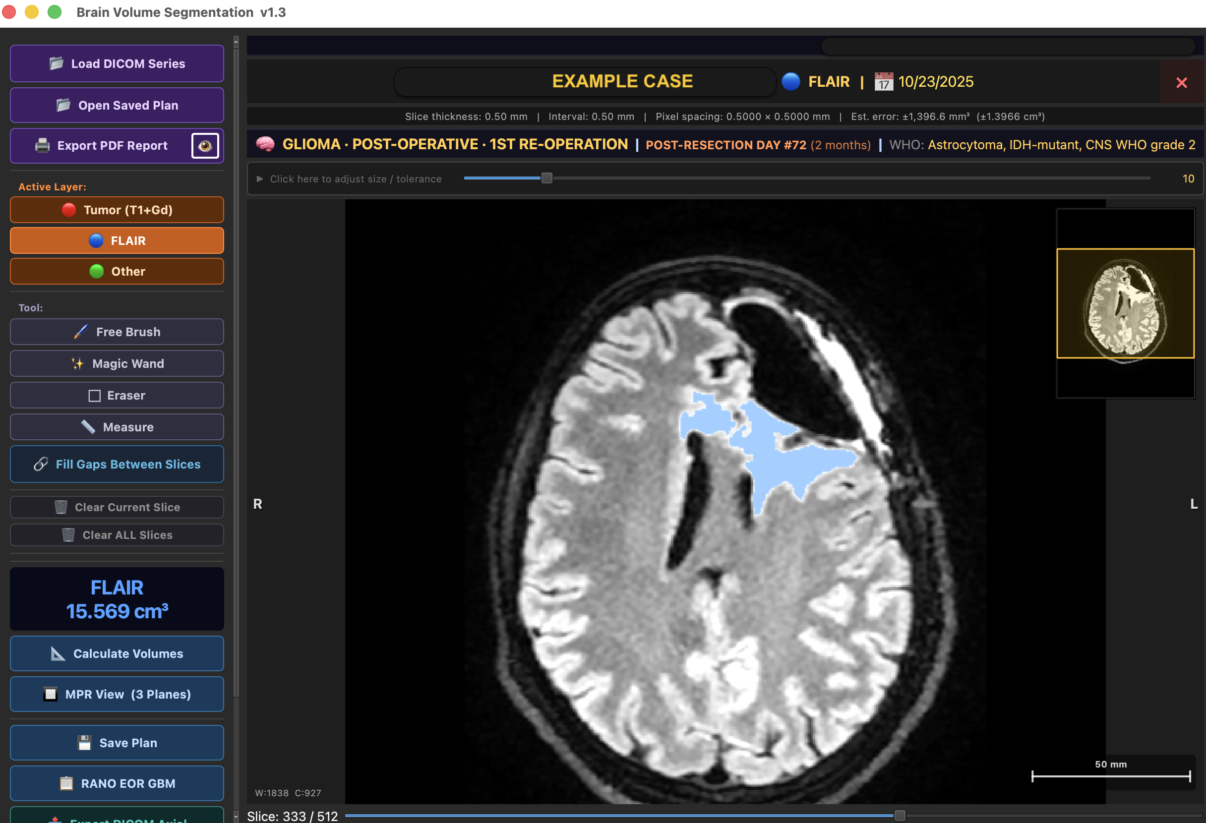
BrainVol app showing FLAIR segmentation of a post-operative glioma case

Designed for clinical and research use

Capabilities

Everything you need for
brain volumetry

From raw DICOM to signed PDF report in under 5 minutes — without leaving your desk.

🪄

Magic Wand Segmentation

Click on a lesion and BrainVol automatically selects the region based on local intensity. Adapts to both CT Hounsfield Units and MRI signal — no manual threshold tuning needed.

CT + MRI
📐

Volumetric Measurement

True voxel-by-voxel volume calculation with partial-volume error estimation. Simultaneous CE and nCE (FLAIR) compartment volumes. ABC/2 comparison for hemorrhage cases.

High accuracy
🧊

3D Reconstruction

One-click 3D surface rendering of any segmented lesion. Rotate, inspect, and include in reports or presentations.

Interactive
🔲

MPR Multiplanar Views

Open coronal and sagittal reconstructions on the fly. Paint and edit the mask directly in any plane, with synchronized window/level matching the axial view.

Axial · Coronal · Sagittal
📊

RANO EOR Classification

Automatic Extent of Resection classification per RANO criteria — gross total, near total, subtotal, or partial resection — computed from CE and FLAIR volumes the moment you segment. Built-in surgical trial reference (MISTIE III, ENRICH, STICH II) for hemorrhage cases.

Gliomas · ICH
📄

PDF Report Generation

Export a professional, print-ready report with axial slice image, volume measurements, patient data, and RANO EOR classification for gliomas. One click.

One click
📈

Follow-up Tracking

Save segmentation plans per patient and compare volumes across time points. Growth percentage with visual trend indicators — ideal for tumor response assessment.

Longitudinal
💾

DICOM Native

Drag and drop any DICOM folder — no conversion needed. Supports MRI (T1+Gd, FLAIR, T2, DWI) and CT. Auto-detects series type and suggests the right layer.

Any scanner
🔬

DICOM Export

Export segmentation masks as RT-STRUCT or Secondary Capture DICOM overlays — compatible with PACS, radiotherapy planning, and research pipelines.

RT-STRUCT · SC
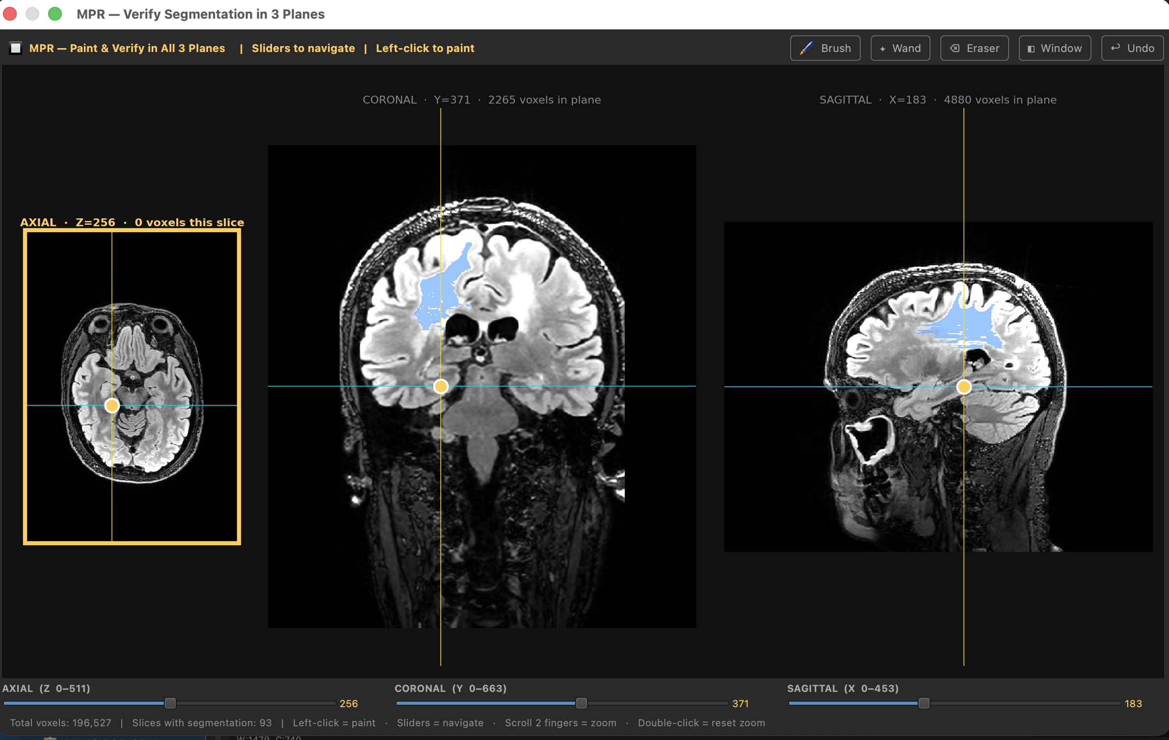
Fine-tune your segmentation
in axial, coronal and sagittal

Every segmentation can be reviewed and retouched in all three planes simultaneously — with Brush, Magic Wand, and Eraser. What you paint in one plane is instantly visible in the other two. No switching apps, no importing masks.

MPR 3-plane segmentation editor
✏️ Brush & Wand 🔄 Instant 3-plane sync 📐 Voxel-accurate

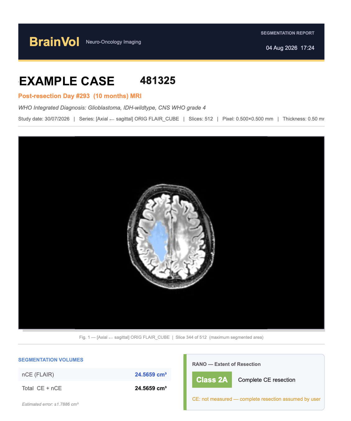
Professional reports,
ready to attach to the clinical file

One click generates a signed PDF with the MRI image, segmentation volumes, RANO Extent of Resection classification, and all study metadata — ready to print or share.

BrainVol PDF segmentation report
Supported Lesions

One tool for every
brain lesion type

Specialized workflows, labels, and clinical criteria for each pathology.

Gliomas
Brain Metastases
Meningiomas
Schwannomas
Intracerebral Hemorrhages (CT)
Subdural Hematomas (CT)
Other
🧠 GLIOMAS
  • ✓ Pre-op / post-op timing
  • ✓ WHO grade classification
  • ✓ CE + nCE (FLAIR) volumes
  • ✓ RANO EOR calculation
🔴 INTRACEREBRAL HEMORRHAGES
  • ✓ Volumetric + ABC/2 estimate
  • ✓ 30 mL surgical threshold
  • ✓ Brain CT window preset
  • ✓ MISTIE III · ENRICH · STICH II
🟤 SUBDURAL HEMATOMAS
  • ✓ Volume > 30 mL threshold
  • ✓ Thickness > 10 mm guidance
  • ✓ Midline shift > 5 mm guidance
  • ✓ CT brain window auto-applied
🟠 BRAIN METASTASES
  • ✓ Multi-lesion segmentation
  • ✓ Per-lesion volume ranking
  • ✓ Total lesion burden
  • ✓ 3D reconstruction per met
Workflow

From DICOM to report
in 4 steps

1

Load your DICOM folder

Drag a folder or use the open dialog. BrainVol scans all series automatically, detects the modality, and suggests the right sequence to load.

2

Select the lesion type

Choose from gliomas, metastases, meningiomas, schwannomas, intracerebral hemorrhages, subdural hematomas, or other. BrainVol configures the right tools and clinical criteria automatically.

3

Segment with Magic Wand or Brush

Click on the lesion with the Magic Wand for automatic region growing, or paint manually with the Brush. Refine with the Eraser. Volume updates live as you paint.

4

Calculate volumes and export

Hit Calculate Volumes for a detailed breakdown — surgical thresholds, RANO EOR, or per-lesion rankings. Export a PDF report in one click.

Demo

See BrainVol in action

Watch a real segmentation from DICOM load to PDF report in under 3 minutes.

Demo video coming soon

Download BrainVol

Available for macOS · Free during beta · Requires activation

🍎 macOS 12 Monterey or later · Apple Silicon + Intel

Requirements: macOS 12+, 8 GB RAM recommended, any DICOM-capable scanner.
Beta version — for research and clinical evaluation purposes.